
Published on
March, 26th, 2025

But first,
let’s cover the basics
Frontal lobe
Cerebellum


Temporal lobe
Occipital lobe
Parietal lobe
Brain stem
Powered by electricity
One way to understand how the brain functions is by measuring its electrical activity. This is done using EEG—short for electroencephalography.


Different ways to measure EEG brain activity. From the more traditional EEG caps to the more consumer friendly EEG wearables.
By placing sensors on the head, we can capture neural activity in real time, down to the millisecond.
Analyzing this data reveals patterns known as brainwaves.
From alpha to gamma:
What are brainwaves?
Brainwaves are patterns of electrical activity in the brain, each occurring at different frequencies: delta, theta, alpha, beta, and gamma.
Frequency refers to how fast a brainwave oscillates. For example, alpha waves cycle 8 to 12 times per second, or 8–12 Hz.
Research on brain activity shows that each brainwave is associated with specific cognitive functions, as outlined below.


Brain maps
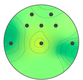
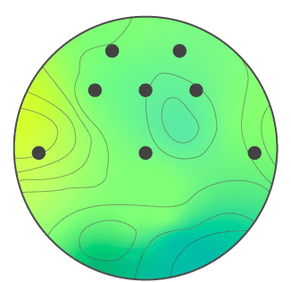
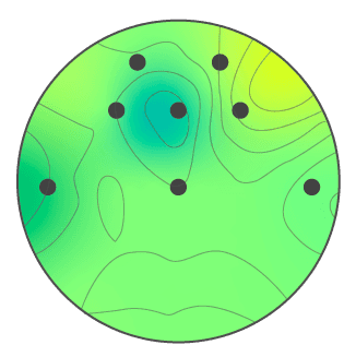
A brainwave strength in a certain region of the brain can tell us about the current mental state, or if seen over time, indicate health issues.
To visualize this, clinicians and researchers use brain maps. These maps can compare a person’s data to a population norm or their own baseline.
Here’s how to read them:


To illustrate this further, here are brain map examples of 4 different mental states. Browse and notice the difference!

BROUGHT TO YOU BY

Measuring brainwaves is great to understand the brain.
Neurofeedback is the way to train it.
View program
What we learned from
EEG recordings
&
brain scans
The female brain shows more activity at faster frequencies
The difference in how our brains are wired
Male and female brainwaves do oscillate differently, but what does that mean and why does it happen?
Here are some reasons why these distinctions occur:
-
Brain structure, connectivity, and blood flow: Females tend to have⁵ ⁶ ⁷increased blood flow⁸
Hormonal Influence: Changes in sex hormones, such as estrogen and progesterone, affect⁹impact areas¹⁰
The brain health indicator, Alpha Peak, differs by sex
What exactly is Alpha Peak?
Try to imagine your brain as a radio, with different channels playing at different frequencies. "Alpha" is a group of channels that play at frequencies between 8 to 12 Hz. The channel that's loudest in that range is what we call Alpha Peak.
A higher Alpha Peak is associated with better cognitive health, processing speed, memory, emotional balance, and the brain's ability to adapt to different mental tasks and challenges.
Brain Frequencies Graph


How does Alpha Peak change with age?

Data recorded from AF7, AF8, TP9, TP10 channels. Total N = 9,644 (Females: N = 4,163; Males: N = 5,481)
Females tend to exhibit higher Alpha Peak values, which appear to reach their maximum later in life, around the age of 38. While this could suggest a potential link to reaching the peak of cognitive function at that age, further research is needed to fully understand its significance.
On the other hand, males peak around the age of 30. So, what explains this difference?
Understanding Alpha Peak differences between sexes
Recent studies¹¹
The brain's Default Mode Network (DMN) plays a crucial role in memory consolidation, introspection, and understanding one’s thoughts and emotions.
It has been suggested¹²
Hormonal changes can influence¹³ ¹⁴
These differences aren’t just structural or functional: they can also be linked to hormonal effects on brain development. For example, androgens (like testosterone) influence brain development differently than estrogen.
Prenatal exposure to androgens can impact¹⁵ ¹⁶
Generally, exposure to androgens is associated with lower alpha peak frequencies or altered alpha activity patterns compared to females.
Our brains help us cope with aging
Despite the differences, female and male brains have one great thing in common: as we age, our brains adapt in ways that help maintain cognitive abilities. Here we see how brainwaves fluctuate over time to support our brain's cognitive functions.
Delta

Theta

Alpha

Beta

Data recorded from AF7, AF8, TP9, TP10 channels. Total N = 9,644 (Females: N = 4,163; Males: N = 5,481)
Low frequency brainwaves (delta and theta) tend to decrease in strength, and their reduction is often linked¹⁷ ¹⁸
On the other hand, relatively higher-frequency waves, like alpha and beta, seem to increase with age.
This increase could be¹⁹
For example, stronger alpha waves might help²⁰
The aging brain compensates for structural changes
Even though the brain undergoes physical changes with age, such as shrinkage in certain areas, reduced connectivity between brain cells, and thinning of the outer layer, cognitive abilities don't always decline²¹ ²²
These adaptations can involve the brain using different regions more effectively or creating new connections to compensate for any decline.
For example, older adults often recruit additional brain regions or show increased activations, which helps²³
Rethinking how we approach brain health
Understanding how our brains stay functional as we age reveals new ways to support cognitive resilience and mental performance. Meanwhile, recognizing the differences between male and female brains opens the door to more tailored approaches to stress management, emotional regulation, and mental health.
By combining these insights, we can create refined solutions that benefit everyone in ways we're just beginning to explore.
Call for researchers
We invite researchers to collaborate with us in uncovering new insights and deepening our understanding of the brain.
Interested in contributing? Send your proposal to research@myndlift.com
References
1) Scheeringa, R., Bastiaansen, M. C. M., Petersson, K. M., Oostenveld, R., Norris, D. G., & Hagoort, P. (2008). Frontal theta EEG activity correlates negatively with the default mode network in resting state. International Journal of Psychophysiology, 67(3), 242–251.
https://doi.org/10.1016/j.ijpsycho.2007.05.017
2) Kim, Y.-W., Kim, S., Jin, M. J., Im, C.-H., & Lee, S.-H. (2024). The Importance of Low-frequency Alpha (8−10 Hz) Waves and Default Mode Network in Behavioral Inhibition. Clinical Psychopharmacology and Neuroscience, 22(1), 53–66.
3) Benchenane, K., Tiesinga, P. H., & Battaglia, F. P. (2011). Oscillations in the prefrontal cortex: A gateway to memory and attention. Current Opinion in Neurobiology, 21(3), 475–485. https://doi.org/10.1016/j.conb.2011.01.004
4) Picazio, S., Veniero, D., Ponzo, V., Caltagirone, C., Gross, J., Thut, G., & Koch, G. (2014). Prefrontal Control over Motor Cortex Cycles at Beta Frequency during Movement Inhibition. Current Biology, 24(24), 2940–2945.
5) Serio, B., Hettwer, M. D., Wiersch, L., Bignardi, G., Sacher, J., Weis, S., Eickhoff, S. B., & Valk, S. L. (2024). Sex differences in functional cortical organization reflect differences in network topology rather than cortical morphometry. Nature Communications, 15(1), 7714.
https://doi.org/10.1038/s41467-024-51942-1
6) Makwana, B., Tart-Zelvin, A., Xu, X., Gunstad, J. J., Cote, D. M., Poppas, A., Cohen, R. A., & Sweet, L. H. (2020). Cerebrovascular Perfusion among Older Adults with and without Cardiovascular Disease. Journal of Neuroimaging : Official Journal of the American Society of Neuroimaging, 30(6), 851–856. https://doi.org/10.1111/jon.12757
7) Gong, G., He, Y., & Evans, A. C. (2011). Brain Connectivity: Gender Makes a Difference. The Neuroscientist, 17(5), 575–591.
8) Weissman‐Fogel, I., Moayedi, M., Taylor, K. S., Pope, G., & Davis, K. D. (2010). Cognitive and default‐mode resting state networks: Do male and female brains “rest” differently? Human Brain Mapping, 31(11), 1713–1726.
9) Styne, D., Grumbach, M., Dennis, M., Styne, J., & Melvin, M. (1998). Puberty: Ontogeny, neuroendocrinology, physiology, and disorders.
10) Woolley, C. S., & McEwen, B. S. (1994). Estradiol regulates hippocampal dendritic spine density via an N-methyl-D-aspartate receptor-dependent mechanism. The Journal of Neuroscience: The Official Journal of the Society for Neuroscience, 14(12), 7680–7687.
11) Lombardo, M., Auyeung, B., Pramparo, T., Quartier, A., Courraud, J., Holt, R& Baron‐Cohen, S. (2018). Sex-specific impact of prenatal androgens on social brain default mode subsystems. Molecular Psychiatry, 25(9), 2175-2188.
12) Lombardo, M., Auyeung, B., Pramparo, T., Quartier, A., Courraud, J., Holt, R& Baron‐Cohen, S. (2018). Sex-specific impact of prenatal androgens on social brain default mode subsystems. Molecular Psychiatry, 25(9), 2175-2188.
13) Lombardo, M., Auyeung, B., Pramparo, T., Quartier, A., Courraud, J., Holt, R& Baron‐Cohen, S. (2018). Sex-specific impact of prenatal androgens on social brain default mode subsystems. Molecular Psychiatry, 25(9), 2175-2188.
https://doi.org/10.1038/s41380-018-0198-y
14) T. De Bondt, et al.
Stability of resting state networks in the female brain during hormonal changes and their relation to premenstrual symptoms Brain Res., 1624 (2015), pp. 275-285
15) Andreano, J. M., Touroutoglou, A., Dickerson, B., & Barrett, L. F. (2018). Hormonal Cycles, Brain Network Connectivity, and Windows of Vulnerability to Affective Disorder. Trends in Neurosciences, 41(10), 660–676.
https://doi.org/10.1016/j.tins.2018.08.007
16) J. Engman, et al.
Hormonal cycle and contraceptive effects on amygdala and salience resting-state networks in women with previous affective side effects on the pill Neuropsychopharmacology, 43 (2018), pp. 555-563
17) Karakaş, S. (2020). A review of theta oscillation and its functional correlates. International Journal of Psychophysiology, 157, 82–99.
https://doi.org/10.1016/j.ijpsycho.2020.04.008
18) Siwek, M. E., Müller, R., Henseler, C., Trog, A., Lundt, A., Wormuth, C., Broich, K., Ehninger, D., Weiergräber, M., & Papazoglou, A. (2015). Altered Theta Oscillations and Aberrant Cortical Excitatory Activity in the 5XFAD Model of Alzheimer’s Disease. Neural Plasticity, 2015(1), 781731. https://doi.org/10.1155/2015/781731
19) Penhale, S. H., Arif, Y., Schantell, M., Johnson, H. J., Willett, M. P., Okelberry, H. J., Meehan, C. E., Heinrichs‐Graham, E., & Wilson, T. W. (2024). Healthy aging alters the oscillatory dynamics and fronto‐parietal connectivity serving fluid intelligence. Human Brain Mapping, 45(3). https://doi.org/10.1002/hbm.26591
20) Springer, S. D., Okelberry, H. J., Willett, M. P., Johnson, H. J., Meehan, C. E., Schantell, M., Embury, C. M., Rempe, M. P., & Wilson, T. W. (2023). Age-related alterations in the oscillatory dynamics serving verbal working memory processing. Aging, 15(24), 14574–14590.
21) Martins, R., Joanette, Y., & Monchi, O. (2015). The implications of age-related neurofunctional compensatory mechanisms in executive function and language processing including the new Temporal Hypothesis for Compensation. Frontiers in Human Neuroscience, 9. https://doi.org/10.3389/fnhum.2015.00221
22) Behfar, Q., Behfar, S. K., von Reutern, B., Richter, N., Dronse, J., Fassbender, R., Fink, G. R., & Onur, O. A. (2020). Graph Theory Analysis Reveals Resting-State Compensatory Mechanisms in Healthy Aging and Prodromal Alzheimer’s Disease. Frontiers in Aging Neuroscience, 12. https://doi.org/10.3389/fnagi.2020.576627
23) Behfar, Q., Behfar, S. K., von Reutern, B., Richter, N., Dronse, J., Fassbender, R., Fink, G. R., & Onur, O. A. (2020). Graph Theory Analysis Reveals Resting-State Compensatory Mechanisms in Healthy Aging and Prodromal Alzheimer’s Disease. Frontiers in Aging Neuroscience, 12. https://doi.org/10.3389/fnagi.2020.576627
